Expansion Microscopy on Saccharomyces cerevisiae
Paper published in microPublication Biology, January 2022
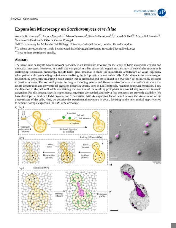
The manuscript describes two distinct protocols: one for preparing coverslips for microscopy analysis of yeast cells, and the other for performing Expansion Microscopy (ExM) on Saccharomyces cerevisiae. In the first protocol, coverslips are cleaned, coated, and cells are mounted on them for observation. The cells are grown, fixed, digested, and embedded in agarose gels. In the second protocol, slides are prepared, denatured, expanded, and stained for ExM. The authors have developed a modified ExM protocol for S. cerevisiae, which includes the use of zymolyase for cell wall digestion and DAPI and NHS-Ester BODIPY FL for staining. The manuscript also discusses the importance of assessing specimen integrity and expansion factor during the experimental process.