Technology
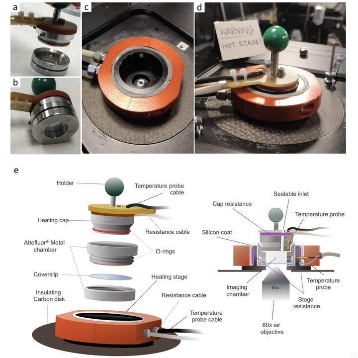
The sulfoscope is a novel microscopy platform developed by Pulschen et al. that enables live-cell imaging of hyperthermophilic archaea like Sulfolobus acidocaldarius. These organisms have optimal growth temperatures of 70-80°C, making it challenging to image them while maintaining physiological growth conditions. To overcome this barrier, the authors designed a heated imaging chamber called the sulfoscope that can stably maintain high temperatures for extended periods of time.
The key innovation of the sulfoscope is the integration of two independent heating elements - a heated cap and a heated stage. The imaging chamber itself sits in between these two heating elements. The cap and stage can be heated to different target temperatures, with the cap set 5°C higher than the stage. This temperature gradient helps minimize thermal gradients across the imaging chamber and prevent excessive water evaporation over long experiments. The entire setup is built to tightly fit standard inverted microscope bodies.
The heated cap is constructed out of metal and contains electric heating elements and insulation. It can be sealed to the imaging chamber using an O-ring to prevent condensation of evaporated water on the underside of the cap. The custom-built heated stage is similarly constructed and designed to perfectly fit the imaging chamber. Together, the dual heating elements enable the chamber temperature to quickly reach equilibrium and remain stable within 0.5°C for the duration of experiments.
To enable live imaging, S. acidocaldarius cells are pre-labeled with thermostable fluorescent probes like Nile Red and SYBR Safe. The cells are then immobilized under a heated Gelrite pad in the sulfoscope chamber. The Gelrite pad gently restricts cell movement while buffering against thermal shock. Multiple aspects of the protocol are optimized to minimize heat loss during transport and setup - from pre-heating hardware to rapidly transferring cell cultures.
With this system, the authors are able to continuously image S. acidocaldarius cells dividing under native growth conditions up to 75°C. They observe the dynamics of coordinated DNA reorganization, compaction, and segregation coupled to cytokinetic furrowing. The cells initially transition from a diffuse DNA state to semi-compact segregated nucleoids before undergoing chromosome condensation coincident with division furrowing. Quantitatively similar dynamics are seen with and without Gelrite immobilization, indicating the natural sequence of events.
By deleting different ESCRT-III components involved in cell division, Pulschen et al. also uncover distinct functional roles for the CdvB1 and CdvB2 proteins using live-cell imaging. While loss of CdvB1 causes occasional division failure, loss of CdvB2 primarily disrupts division symmetry. Further analysis suggests CdvB2 helps prevent slippage of the cytokinetic ring, whereas CdvB1 aids constriction.
The sulfoscope enables live-cell imaging of thermophilic organisms like S. acidocaldarius by maintaining high constant temperatures. It reveals tight coupling between DNA reorganization and cell division events. By expanding our view into archaeal cell biology, the system further highlights functional differences between division proteins previously thought to be interchangeable. The sulfoscope platform and approach open up new possibilities for studying dynamic processes in thermophiles.
Publications featuring Sulfoscope
|
Live imaging of a hyperthermophilic archaeon reveals distinct roles for two ESCRT-III homologs in ensuring a robust and symmetric division Andre Arashiro Pulschen, Delyan R Mutavchiev, Siân Culley, Kim Nadine Sebastian, Jacques Roubinet, Marc Roubinet, Gabriel Tarrason Risa, Marleen van Wolferen, Chantal Roubinet, Uwe Schmidt, Gautam Dey, Sonja-Verena Albers, Ricardo Henriques, Buzz Baum Paper published in Current Biology, January 2020 Technologies: Sulfoscope Funded by: Wellcome Trust DOI: 10.1016/j.cub.2020.05.021 |
|