An accessible framework to achieve multi-dimensional live-cell super-resolution high-content screening
Type: Tools and Resources Development Fund
Principal Investigator: Ricardo Henriques
Investigators: Ricardo Henriques, Robin Ketteler
Start-date: December 2017
End-date: December 2018
Grant Code: BB/P027431/1
Abstract Imagine we could see how the molecular machineries inside cells behave, form networks and develop into complex tissues! What has been a dream for humanity for centuries has become ever closer to possibility through multiple technical advances. This development started with the optical microscope in the 17th century that enabled the visualisation of cells and bacteria, confocal and electron microscopy in the 1950's to visualise smaller structures, and culminated in the advent of super-resolution microscope in the 2000's that now enable us to see specific protein complexes at high resolution.
Microscopy has been a ground-breaking technology that most academic labs and pharma companies make use of to assay drugs and visualise disease models. One limitation of such techniques has been the time required for imaging, which made it impractical to acquire a large number of images - a pre-requisite for screening thousands of drugs. Therefore, high-content imaging platforms have been developed that enable the acquisition of thousands of images in a relatively short time frame. Yet, images generated by high-content screening microscopes often lack the resolution, for instance for visualising small sub-cellular structures. Here, in this proposal we aim to combine two methods in order to generate the first super-resolution high-content screening platform (SR-HCS). This platform will truly transform research in cell biology and enable discoveries at an unprecedented speed and resolution.
Technical Summary High content biology has revolutionised our understanding of biological systems. The particular application of image-based high-content screening (HCS) essays into cell biology has provided us critical new insights into the role of gene networks, stochasticity in cell population behaviour, and the discovery of how tailored compounds modulate normal cell behaviour or in the presence of disease, among others. Chiefly, HCS brings a big data perspective into cell biology, allowing the image analysis of the behaviour of large cell populations under different states and conditions. However, to date, HCS still relies on underlying conventional imaging approaches such as confocal or widefield microscopy, resolution limited to ~500nm. This limit implies that HCS cannot accurately resolve the small details of molecular events critically regulating cell dynamics, such as molecular interactions and the formation of supra-molecular complexes existing at scales of few nanometres.
There is an urgent need to develop higher resolution instruments in high-content cell biology. For example, several cellular structures become only visible when examining with super-resolution microscopy. Typical image analysis workflows struggle to separate objects when looking at small intracellular organelles. Two technical advances in the last years have made it possible to overcome this limitation and can provide high resolution images on a HCS platform: 1) the development of a spinning disk confocal HCS platform (e.g. the PerkinElmer Opera Phenix), and 2) the development of image analysis software that enables the post-acquisition resolution of images to achieve sub-diffraction limit values (e.g. SRRF). Here, we aim to combine these two techniques and to develop the first super-resolution high-content screening platform (SR-HCS), in collaboration with PerkinElmer. We anticipate that this unique system will enable breakthrough discoveries in fundamental cell biology and biomedical research.
Planned Impact Academic: We aim to deliver to researchers a novel large-scale imaging framework to study cell behaviour at the nanoscale. This super-resolution high-content screening platform will be the first such platform world-wide and enable academics, as well as interested pharmaceutical partners, to benefit from this technology. We expect that any lab engaged in biological or biomedical imaging to adopt this technology, in particular those that are interested in the study of sub-cellular organelles and protein complexes. It will allow findings from a novel imaging approach to be made available, enabling new avenues of interdisciplinary collaborative.
Commercial: It will be possible to rapidly translate the analytical and imaging developments from this project into applications of interest to biotechnology, microscopy and imaging companies. The data generated will be of high-value to the pharma industry as it has the potential to identify fundamental cell behaviour in the presence of disease, allowing for the tailored design of new drug targets. Most importantly, we are closely collaborating with PerkinElmer in this project to ensure that our software and hardware developments can be translated into a potential commercial solution.
Training: The PDRA to be recruited will receive continuous cross-disciplinary training and mentoring, which will aid his/her career progression. The skills learned over the course of this project, namely image analysis and high-content screening, are very rare to find in the UK and in high demand. We expect that training of researchers skilled in this area will contribute immensely to the UK competitiveness in these areas, both in academia and industry. In addition, the PDRA will benefit from generic skills gained on training courses at UCL, through co-authoring papers, grants and reviews and by presenting this work at international meetings. The project described here will be ideal for introducing students from different backgrounds to interdisciplinary research in life and physical sciences. Moreover, a large number of students will benefit from involvement in this interdisciplinary level research through rotation projects, MRes and tutorial activities associated with these programmes. Similarly, undergraduates will be exposed to this work through internships and short projects. Finally, by publicising this work, we expect to attract better and brighter students and researchers to the UK.
General Societal and Economical Impact: Microscopy approaches and instrumentation is one of the fastest growing areas in biomedical research, critical not only for cell biology, but for the direct discovery of new molecular targets mitigating human disease. Over the past decades, the UK has been a world leader in these developments. This project will enable and supply new approaches crucial for enhancing microscopy and cell biology studies, supplying UK research with novel state-of-the-art and experimental methods - a key attractor for industrial R&D collaborations - and empowering research in the UK with some of the most advanced imaging facilities in Europe. Locally, UCL (where this project will be housed) has made a commitment to make the interface between medicine, biophysics and imaging a key priority, contributing to the long-term sustainability of these studies and serving as a seeding source for collaborations with our partners such as the new Francis Crick Institute and NHS Hospitals (e.g. Royal Free Hospital).
Outreach: RH and RK have been involved in interactions with the wider community through media appearances, public discussions and school visits. Through this type of outreach we expect this work to reach a wide audience; giving the public a better understanding of how next-generation imaging technology may impact research, health and disease. Moreover, through our involvement in EMBO, EU and UK-South Africa networks, we expect this work to reach the global scientific community.
Supported publications

|
Nanoscale colocalization of NK cell activating and inhibitory receptors controls signal integration David Tomaz, Pedro Matos Pereira, Nadia Guerra, Julian Dyson, Keith Gould, Ricardo Henriques Paper published in Frontiers in Immunology, January 2022 Technologies: NanoJ () and NanoJ-SQUIRREL () Funded by: BBSRC, EMBO and ERC DOI: 10.3389/fimmu.2022.868496 |
|
|
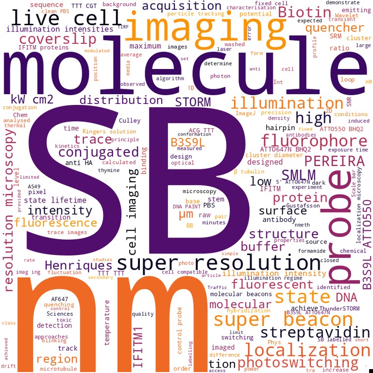
Super‐beacons - Open‐source probes with spontaneous tuneable blinking compatible with live‐cell super‐resolution microscopy Pedro M Pereira, Nils Gustafsson, Mark Marsh, Musa M Mhlanga, Ricardo Henriques Paper published in Traffic, January 2020 Technologies: NanoJ (), NanoJ-Fluidics (), NanoJ-SQUIRREL (), NanoJ-SRRF () and Super-Beacons Funded by: BBSRC and Wellcome Trust DOI: 10.1111/tra.12728 |
|
|
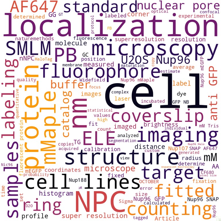
Nuclear pores as versatile reference standards for quantitative superresolution microscopy Jervis Vermal Thevathasan, Maurice Kahnwald, Konstanty Cieśliński, Philipp Hoess, Sudheer Kumar Peneti, Manuel Reitberger, Daniel Heid, Krishna Chaitanya Kasuba, Sarah Janice Hoerner, Yiming Li, Yu-Le Wu, Markus Mund, Ulf Matti, Pedro Matos Pereira, Ricardo Henriques, Bianca Nijmeijer, Moritz Kueblbeck, Vilma Jimenez Sabinina, Jan Ellenberg, Jonas Ries Paper published in Nature Methods, September 2019 Technologies: CARE (), NanoJ (), NanoJ-SQUIRREL (), NanoJ-SRRF () and Nuclear-Pores as references Funded by: BBSRC and Wellcome Trust News: Mirage News DOI: 10.1038/s41592-019-0574-9 |
|
|
|
Artificial intelligence for microscopy - what you should know Lucas von Chamier, Romain F. Laine, Ricardo Henriques Review published in Biochemical Society Transactions, July 2019 Technologies: CARE (), NanoJ (), NanoJ-Fluidics (), NanoJ-SQUIRREL () and NanoJ-SRRF () Funded by: BBSRC and Wellcome Trust News: Azooptics.com DOI: 10.1042/bst20180391 |
|

|
Nanoscale polarization of the entry fusion complex of vaccinia virus drives efficient fusion Robert D. M. Gray, David Albrecht, Corina Beerli, Moona Huttunen, Gary H. Cohen, Ian J. White, Jemima J. Burden, Ricardo Henriques, Jason Mercer Paper published in Nature Microbiology, July 2019 Technologies: NanoJ () and NanoJ-VirusMapper Funded by: BBSRC and Wellcome Trust Blogs: Nature Microbiology Community DOI: 10.1038/s41564-019-0488-4 |
|

|
Automating multimodal microscopy with NanoJ-Fluidics Pedro Almada, Pedro M. Pereira, Siân Culley, Ghislaine Caillol, Fanny Boroni-Rueda, Christina L. Dix, Guillaume Charras, Buzz Baum, Romain F. Laine, Christophe Leterrier, Ricardo Henriques Paper published in Nature Communications, March 2019 Technologies: NanoJ (), NanoJ-Fluidics (), NanoJ-SQUIRREL (), NanoJ-SRRF () and NanoJ-VirusMapper Funded by: BBSRC and Wellcome Trust News: Technology Times, MSN, DNYUZ and Express Informer DOI: 10.1038/s41467-019-09231-9 |
|
|
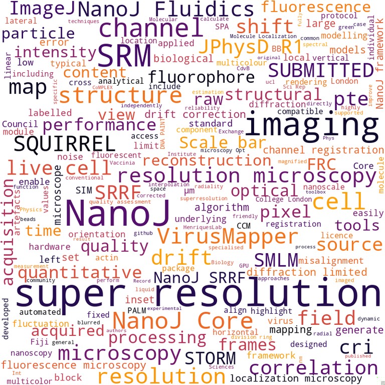
NanoJ - a high-performance open-source super-resolution microscopy toolbox Romain F Laine, Kalina L Tosheva, Nils Gustafsson, Robert D M Gray, Pedro Almada, David Albrecht, Gabriel T Risa, Fredrik Hurtig, Ann-Christin Lindås, Buzz Baum, Jason Mercer, Christophe Leterrier, Pedro M Pereira, Siân Culley, Ricardo Henriques Paper published in Journal of Physics D - Applied Physics, January 2019 Technologies: CARE (), NanoJ (), NanoJ-SQUIRREL (), NanoJ-SRRF (), NanoJ-VirusMapper and QuickPALM Funded by: BBSRC and Wellcome Trust DOI: 10.1088/1361-6463/ab0261 |
|
|
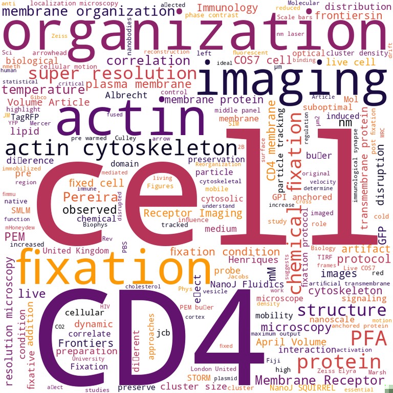
Fix your membrane receptor imaging - actin cytoskeleton and CD4 membrane organization disruption by chemical fixation Pedro M Pereira, David Albrecht, Siân Culley, Caron Jacobs, Mark Marsh, Jason Mercer, Ricardo Henriques Published in Frontiers in immunology, January 2019 Technologies: NanoJ (), NanoJ-Fluidics () and NanoJ-SQUIRREL () Funded by: BBSRC and Wellcome Trust DOI: 10.3389/fimmu.2019.00675 |
|
|
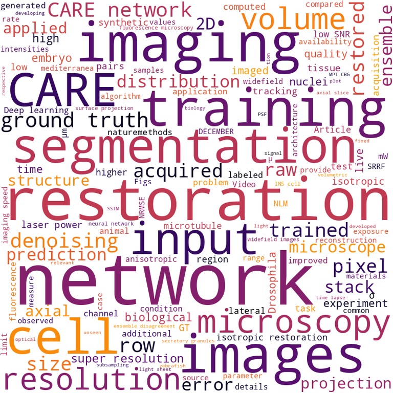
Content-aware image restoration - pushing the limits of fluorescence microscopy Martin Weigert, Uwe Schmidt, Tobias Boothe, Andreas Müller, Alexandr Dibrov, Akanksha Jain, Benjamin Wilhelm, Deborah Schmidt, Coleman Broaddus, Siân Culley, Mauricio Rocha-Martins, Fabián Segovia-Miranda, Caren Norden, Ricardo Henriques, Marino Zerial, Michele Solimena, Jochen Rink, Pavel Tomancak, Loic Royer, Florian Jug, Eugene W. Myers Paper published in Nature Methods, November 2018 Technologies: CARE (), NanoJ-SQUIRREL () and NanoJ-SRRF () Funded by: BBSRC and Wellcome Trust News: Technology Networks, VBIO, Innovations Report and Informationsdienst Wissenschaft DOI: 10.1038/s41592-018-0216-7 |
|
|
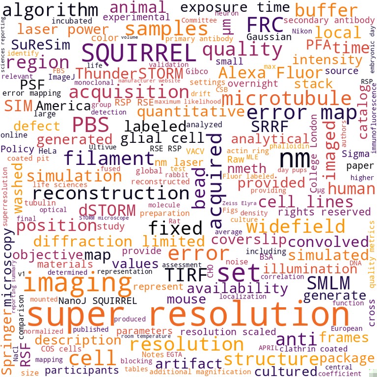
Quantitative mapping and minimization of super-resolution optical imaging artifacts Siân Culley, David Albrecht, Caron Jacobs, Pedro Matos Pereira, Christophe Leterrier, Jason Mercer, Ricardo Henriques Paper published in Nature Methods, February 2018 Technologies: NanoJ-SQUIRREL (), NanoJ-SRRF () and QuickPALM Funded by: BBSRC and Wellcome Trust News: physicsworld.com DOI: 10.1038/nmeth.4605 |
|
|
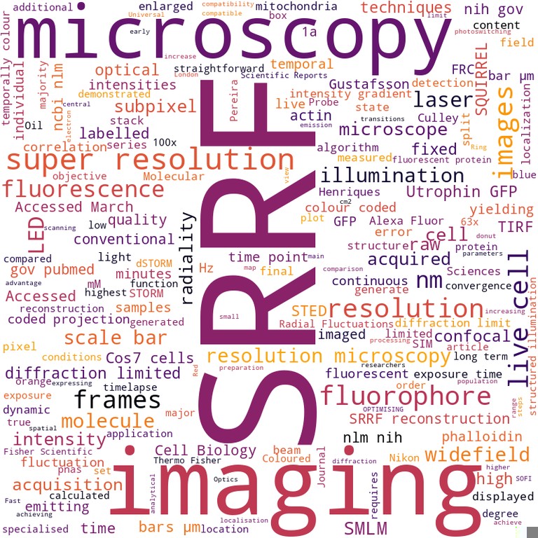
SRRF - Universal live-cell super-resolution microscopy Siân Culley, Kalina L Tosheva, Pedro Matos Pereira, Ricardo Henriques Paper published in The international journal of biochemistry & cell biology, January 2018 Technologies: NanoJ-SQUIRREL () and NanoJ-SRRF () Funded by: BBSRC and Wellcome Trust DOI: 10.1016/j.biocel.2018.05.014 |
|

|
Septins recognize and entrap dividing bacterial cells for delivery to lysosomes Sina Krokowski, Damian Lobato-Marquez, Arnaud Chastanet, Pedro Matos Pereira, Dimitrios Angelis, Dieter Galea, Gerald Larrouy-Maumus, Ricardo Henriques, Elias T Spiliotis, Rut Carballido-Lopez, Serge Mostowy Published in Cell host & microbe, January 2018 Technologies: NanoJ-VirusMapper Funded by: BBSRC DOI: 10.1016/j.chom.2018.11.005 |
|
News
- 2024-05-13: News outlet Azooptics.com highlights Chamier et al. Biochemical Society Transactions 2019 [external link]
- 2022-11-15: News outlet Technology Times highlights Almada et al. Nature Communications 2019 [external link]
- 2022-06-18: News outlet MSN highlights Almada et al. Nature Communications 2019 [external link]
- 2022-06-18: News outlet DNYUZ highlights Almada et al. Nature Communications 2019 [external link]
- 2022-06-18: News outlet Express Informer highlights Almada et al. Nature Communications 2019 [external link]
- 2021-07-14: News outlet Technology Networks highlights Weigert et al. Nature Methods 2018 [external link]
- 2020-10-27: News outlet Mirage News highlights Thevathasan et al. Nature Methods 2019 [external link]
- 2019-08-19: News outlet This Week in Virology (TWiV) highlights Gray et al. Nature Microbiology 2019 [external link]
- 2019-07-10: Blog Nature Microbiology Community highlights Gray et al. Nature Microbiology 2019 [external link]
- 2018-12-10: News outlet VBIO highlights Weigert et al. Nature Methods 2018 [external link]
- 2018-12-07: News outlet Innovations Report highlights Weigert et al. Nature Methods 2018 [external link]
- 2018-12-07: News outlet Informationsdienst Wissenschaft highlights Weigert et al. Nature Methods 2018 [external link]
- 2018-05-14: News outlet physicsworld.com highlights Culley et al. Nature Methods 2018 [external link]