Enabling Live-Cell Super Resolution Imaging Through Lattice Light Sheet Microscopy
Type: ALERT
Principal Investigator: Franck Pichaud
Investigators: Franck Pichaud, Karl Matter, Alan Lowe, Guillaume Charras, Ricardo Henriques, Buzz Baum
Start-date: January 2017
End-date: May 2018
Grant Code: BB/R000697/1
Abstract The aim with this application is to set up a world-leading pole of excellence for live-cell Super-Resolution imaging. The cell is the fundamental unit of all complex life forms and so understanding the biology of the cell and its component parts is a key goal in life science and medicine. Next-generation microscopes are profoundly changing the way cells, sub-cellular structures, and tissues can be studied. More than ever before, our ability to remain a leading research institution in biomedical sciences requires us to stay abreast of new, transformative imaging technologies. To further our understanding of health and diseases requires a deep knowledge of the cell and its constituents. One main path toward this goal is to be able to image the behavior and functional properties of large populations of cells over time. This must be done at high spatial and temporal resolution so that dynamic processes can be discovered and studied. Importantly, imaging of cells and tissues must also be done while preventing photo-toxicity, which can lead to artefacts and even cell death. Advanced optical techniques such as light sheet microscopy, allow for such dynamic studies at low photo-toxicity. Here we request funds to enable us to acquire and set up a novel ultra-fast, exquisitely sensitive, lattice light sheet microscope that will transform our research capabilities in cell and developmental biology. The microscope will be accessible to a wide variety of researchers addressing many outstanding questions relating to embryonic development, cell biology and neurobiology in both normal and disease conditions.
To accurately understand and study cellular systems requires imaging at low-illumination minimizing light-induced perturbation of the sample. Photo-toxicity is a main issue in light microscopy, that is know to induce artefacts and cell death. This critical problem is being addressed at UCL with the aim to provide our research-community with the capacity to follow cells at high-resolution and with minimal perturbation. UCL has been instrumental in developing and implementing optical approaches tackling the problem of photo-toxicity. In the recent years, UCL has entered a new and exciting partnership with a small size company, 3i, whose area of expertise is to design and assemble state-of-the-art microscope, including new generating light sheet microscopes which enable live cell imaging with minimum photo-toxicity. Together UCL and 3i are engaged in joint projects that aim at developing new technologies enabling Super-Resolution long-term imaging, through light-sheet microscopy (LLSM). Here we request for support to acquire a LLSM - considered the best performing microscope for simultaneous imaging depth, speed, resolution and specimen viability. This system is uniquely available through 3i and will be transformative for cell and developmental biology at UCL by enabling long-term, Super-Resolution imaging of cells and subcellular structures and compartment. The LLSM will complement another type of light sheet microscope called Dual-View Inverted Selective Plane Illumination Microscope (diSPIM), which will be assembled by 3i at the LMCB as part of our collaborative agreement. This particular combination of instruments will transform the ways UCL researchers are designing and implementing live and tissue imaging.
Technical Summary LLSM relies on the generation of thin sheets of light, perpendicular to the image detection path. These light sheets are optical lattices form by three-dimensional interference patterns. In dithered mode, the LLSM scans these patterns along an axis tangent to the focal plane, allowing for an image to be collected representing a z-plane, with diffraction limit xy-resolution and highly thin (~370nm) z-section illumination when compared to that of other light sheet microscopy method (>1000nm). The advantage of LLSM is it constrains sample illumination in Z to the focal plane of the objective thus minimizing any out-of-focus excitation of fluorophores - this allows for a considerably decrease in sample illumination while providing a high detection signal-to-noise ratio.
Additionally, LLSM achieves super-resolution through structured illumination microscopy (SIM) by discretely stepping the formed lattice over the sample, effectively increasing the resolution of the system by 2-folds. The combination of LLSM, diSPIM and next-generation optical approaches being implemented at UCL such as TIRF-SIM, STED, SMLMS (e.g.: PALM, STORM, SRRF), SPIM and OPT, will generate an exceptional internationally recognized research environment for fundamental cell biology research. As part of this application, the LLSM will be set up and used to study in details three main area in cell and developmental biology: mitosis, migration and polarity. LLSM will allow for Super-Resolution, live imaging of spindle assembly and its relationship to the cortex and Golgi apparatus, which are al structures that a prone to light induced damage. LLSM will also permit to study epithelial cell polarization and in particular the actomyosin network and apical cortex, which drive plasma membrane specialization at unprecedented high time resolution whilst achieving super-resolution in living cells.
Planned Impact UCL has an excellent track record of multi-institution research - as example, ~20% of publications using the recently formed UCL SuRF are from research lead by groups outside of UCL. Promoting these engagements is a major priority at UCL, as the benefits of sharing advanced resources across research institutions primes the capacity for outstanding research.
Dissemination of findings to the scientific community: will be through publication in international peer-reviewed scientific journals. All PI in the application are leaders in their fields and are frequently invited to present their work at international meetings. In addition, user groups that meet regularly at UCL and nearby universities, such as the Physics of Living Matter meetings, and the "London Super-Resolution Group" (LSR-co-organized by Ricardo Henrique), offer ideal forums to update the national and international community on advances and opportunities. Thus, the results emerging from our work will have impact far beyond our immediate circle of specialists.
Dissemination of findings to the end user: The LLSM will be incorporated in our exciting SuRF imaging platform at the LMCB (https://www.ucl.ac.uk/super-resolution) and will also be featured on the Bloomsbury Consortium for Advanced Light Microscopy, B_CALM@UCL web site (http://www.ucl.ac.uk/advanced-light-microscopy) through which information on resources, applications, etc. will be made available. When appropriate, information will be released through the UCL or BBSRC Press Offices. UCL operates a schools engagement programme to encourage young students to develop an interest in cell biology. Students will be able to visit the facility, see the instruments in use and view new findings.
Establish collaborations within the scientific community and with industry: we will directly link bioscience researchers and instrument developers (such as our partner 3i) to provide a more integrated approach to Super Resolution live-cell imaging. We will hold regular (at 3-6 month intervals) user meetings where results will be presented and discussed. Commercial opportunities that will arise from our R&D work on LLSM will be taken forward through UCL Business and BBSRC Technology Transfer. LLSM will also enable new opportunities for therapy and drug development. In this respect LLSM and the will complement the recently established LMCB high throughput, high content screening platform and the Super Resolution Imaging platform which is now associated to the new MRC dementia centre at UCL.
Training of people: UCL hosts a number of Postgraduate Training Programmes including the LMCB MRC funded four year graduate programme, the BBSRC London Interdisciplinary Biosciences PhD Consortium and. CoMPLEX provides a masters training programme that links physics/maths and engineering with biology - much of which is provided by members of the LMCB. Members of the LMCB, notably Ewa Paluch and Buzz Baum, run UCL's new Institute for the Physics of Living Systems, which brings together physicists and biologists, which will include a new Masters programme next year. The new facility will provide opportunities for BBSRC, IPLS and CoMPLEX students to gain insight into advanced imaging methodologies with the view that they may opt to work in this area for rotations and/or thesis projects, providing further opportunities for capacity building and development.
Industrial engagement: through our collaborative agreement with 3i, this proposal will generate a research environment where not only new imaging technologies such as the LLSM drive new research, but feedback from researchers nurses the R&D of novel imaging systems carried by industry. Ultimately, this environment will stimulate not only excellent research but also impact the rapid access to the wide research community of novel biology questions driven imaging hardware development.
Supported publications

|
Nanoscale colocalization of NK cell activating and inhibitory receptors controls signal integration David Tomaz, Pedro Matos Pereira, Nadia Guerra, Julian Dyson, Keith Gould, Ricardo Henriques Paper published in Frontiers in Immunology, January 2022 Technologies: NanoJ () and NanoJ-SQUIRREL () Funded by: BBSRC, EMBO and ERC DOI: 10.3389/fimmu.2022.868496 |
|
|
Between life and death - strategies to reduce phototoxicity in super-resolution microscopy Kalina L Tosheva, Yue Yuan, Pedro Matos Pereira, Siân Culley, Ricardo Henriques Review published in Journal of Physics D - Applied Physics, January 2020 Technologies: CARE (), NanoJ (), NanoJ-Fluidics () and NanoJ-SRRF () Funded by: BBSRC and Wellcome Trust DOI: 10.1088/1361-6463/ab6b95 |
|
|
Super‐beacons - Open‐source probes with spontaneous tuneable blinking compatible with live‐cell super‐resolution microscopy Pedro M Pereira, Nils Gustafsson, Mark Marsh, Musa M Mhlanga, Ricardo Henriques Paper published in Traffic, January 2020 Technologies: NanoJ (), NanoJ-Fluidics (), NanoJ-SQUIRREL (), NanoJ-SRRF () and Super-Beacons Funded by: BBSRC and Wellcome Trust DOI: 10.1111/tra.12728 |
|
|
Nuclear pores as versatile reference standards for quantitative superresolution microscopy Jervis Vermal Thevathasan, Maurice Kahnwald, Konstanty Cieśliński, Philipp Hoess, Sudheer Kumar Peneti, Manuel Reitberger, Daniel Heid, Krishna Chaitanya Kasuba, Sarah Janice Hoerner, Yiming Li, Yu-Le Wu, Markus Mund, Ulf Matti, Pedro Matos Pereira, Ricardo Henriques, Bianca Nijmeijer, Moritz Kueblbeck, Vilma Jimenez Sabinina, Jan Ellenberg, Jonas Ries Paper published in Nature Methods, September 2019 Technologies: CARE (), NanoJ (), NanoJ-SQUIRREL (), NanoJ-SRRF () and Nuclear-Pores as references Funded by: BBSRC and Wellcome Trust News: Mirage News DOI: 10.1038/s41592-019-0574-9 |
|
|
|
Artificial intelligence for microscopy - what you should know Lucas von Chamier, Romain F. Laine, Ricardo Henriques Review published in Biochemical Society Transactions, July 2019 Technologies: CARE (), NanoJ (), NanoJ-Fluidics (), NanoJ-SQUIRREL () and NanoJ-SRRF () Funded by: BBSRC and Wellcome Trust News: Azooptics.com DOI: 10.1042/bst20180391 |
|

|
Nanoscale polarization of the entry fusion complex of vaccinia virus drives efficient fusion Robert D. M. Gray, David Albrecht, Corina Beerli, Moona Huttunen, Gary H. Cohen, Ian J. White, Jemima J. Burden, Ricardo Henriques, Jason Mercer Paper published in Nature Microbiology, July 2019 Technologies: NanoJ () and NanoJ-VirusMapper Funded by: BBSRC and Wellcome Trust Blogs: Nature Microbiology Community DOI: 10.1038/s41564-019-0488-4 |
|
|
Super-resolution fight club - assessment of 2D and 3D single-molecule localization microscopy software Daniel Sage, Thanh-An Pham, Hazen Babcock, Tomas Lukes, Thomas Pengo, Jerry Chao, Ramraj Velmurugan, Alex Herbert, Anurag Agrawal, Silvia Colabrese, Ann Wheeler, Anna Archetti, Bernd Rieger, Raimund Ober, Guy M. Hagen, Jean-Baptiste Sibarita, Jonas Ries, Ricardo Henriques, Michael Unser, Seamus Holden Paper published in Nature Methods, April 2019 Technologies: QuickPALM Funded by: BBSRC DOI: 10.1038/s41592-019-0364-4 |
|
|
NanoJ - a high-performance open-source super-resolution microscopy toolbox Romain F Laine, Kalina L Tosheva, Nils Gustafsson, Robert D M Gray, Pedro Almada, David Albrecht, Gabriel T Risa, Fredrik Hurtig, Ann-Christin Lindås, Buzz Baum, Jason Mercer, Christophe Leterrier, Pedro M Pereira, Siân Culley, Ricardo Henriques Paper published in Journal of Physics D - Applied Physics, January 2019 Technologies: CARE (), NanoJ (), NanoJ-SQUIRREL (), NanoJ-SRRF (), NanoJ-VirusMapper and QuickPALM Funded by: BBSRC and Wellcome Trust DOI: 10.1088/1361-6463/ab0261 |
|
|
Fix your membrane receptor imaging - actin cytoskeleton and CD4 membrane organization disruption by chemical fixation Pedro M Pereira, David Albrecht, Siân Culley, Caron Jacobs, Mark Marsh, Jason Mercer, Ricardo Henriques Published in Frontiers in immunology, January 2019 Technologies: NanoJ (), NanoJ-Fluidics () and NanoJ-SQUIRREL () Funded by: BBSRC and Wellcome Trust DOI: 10.3389/fimmu.2019.00675 |
|
|
Content-aware image restoration - pushing the limits of fluorescence microscopy Martin Weigert, Uwe Schmidt, Tobias Boothe, Andreas Müller, Alexandr Dibrov, Akanksha Jain, Benjamin Wilhelm, Deborah Schmidt, Coleman Broaddus, Siân Culley, Mauricio Rocha-Martins, Fabián Segovia-Miranda, Caren Norden, Ricardo Henriques, Marino Zerial, Michele Solimena, Jochen Rink, Pavel Tomancak, Loic Royer, Florian Jug, Eugene W. Myers Paper published in Nature Methods, November 2018 Technologies: CARE (), NanoJ-SQUIRREL () and NanoJ-SRRF () Funded by: BBSRC and Wellcome Trust News: Technology Networks, VBIO, Innovations Report and Informationsdienst Wissenschaft DOI: 10.1038/s41592-018-0216-7 |
|
|
Quantitative mapping and minimization of super-resolution optical imaging artifacts Siân Culley, David Albrecht, Caron Jacobs, Pedro Matos Pereira, Christophe Leterrier, Jason Mercer, Ricardo Henriques Paper published in Nature Methods, February 2018 Technologies: NanoJ-SQUIRREL (), NanoJ-SRRF () and QuickPALM Funded by: BBSRC and Wellcome Trust News: physicsworld.com DOI: 10.1038/nmeth.4605 |
|
|
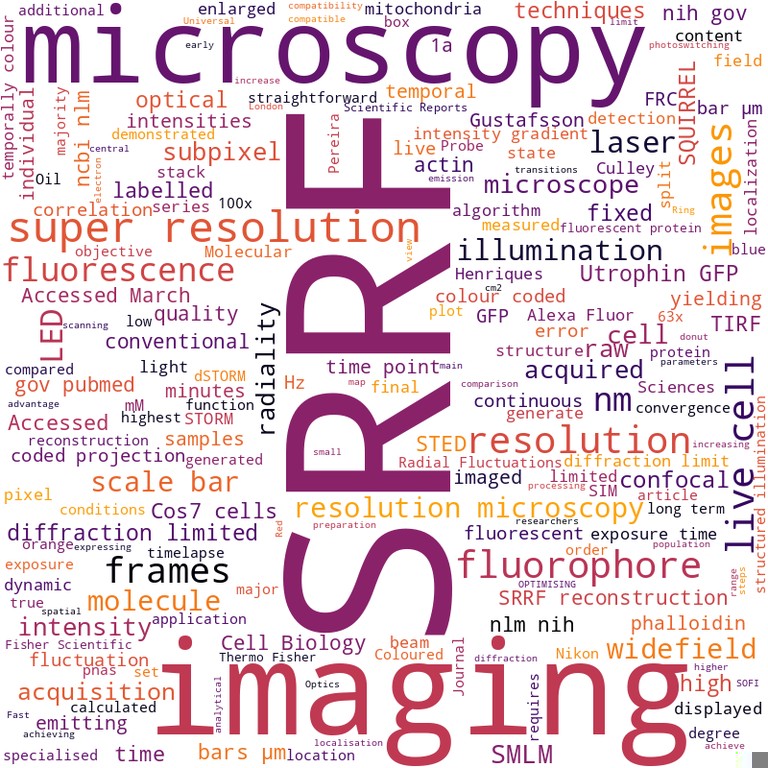
SRRF - Universal live-cell super-resolution microscopy Siân Culley, Kalina L Tosheva, Pedro Matos Pereira, Ricardo Henriques Paper published in The international journal of biochemistry & cell biology, January 2018 Technologies: NanoJ-SQUIRREL () and NanoJ-SRRF () Funded by: BBSRC and Wellcome Trust DOI: 10.1016/j.biocel.2018.05.014 |
|
|
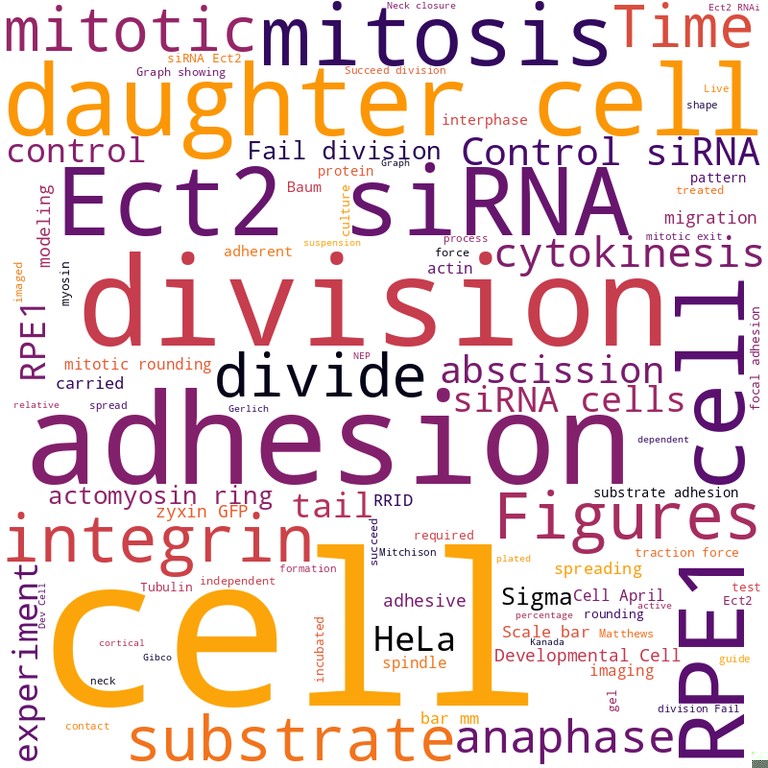
The role of mitotic cell-substrate adhesion re-modeling in animal cell division Christina L Dix, Helen K Matthews, Marina Uroz, Susannah McLaren, Lucie Wolf, Nicholas Heatley, Zaw Win, Pedro Almada, Ricardo Henriques, Michael Boutros, Xavier Trepat, Buzz Baum Published in Developmental cell, January 2018 Funded by: BBSRC DOI: 10.1016/j.devcel.2018.03.009 |
|

|
Septins recognize and entrap dividing bacterial cells for delivery to lysosomes Sina Krokowski, Damian Lobato-Marquez, Arnaud Chastanet, Pedro Matos Pereira, Dimitrios Angelis, Dieter Galea, Gerald Larrouy-Maumus, Ricardo Henriques, Elias T Spiliotis, Rut Carballido-Lopez, Serge Mostowy Published in Cell host & microbe, January 2018 Technologies: NanoJ-VirusMapper Funded by: BBSRC DOI: 10.1016/j.chom.2018.11.005 |
|
News
- 2024-05-13: News outlet Azooptics.com highlights Chamier et al. Biochemical Society Transactions 2019 [external link]
- 2021-07-14: News outlet Technology Networks highlights Weigert et al. Nature Methods 2018 [external link]
- 2020-10-27: News outlet Mirage News highlights Thevathasan et al. Nature Methods 2019 [external link]
- 2019-08-19: News outlet This Week in Virology (TWiV) highlights Gray et al. Nature Microbiology 2019 [external link]
- 2019-07-10: Blog Nature Microbiology Community highlights Gray et al. Nature Microbiology 2019 [external link]
- 2018-12-10: News outlet VBIO highlights Weigert et al. Nature Methods 2018 [external link]
- 2018-12-07: News outlet Innovations Report highlights Weigert et al. Nature Methods 2018 [external link]
- 2018-12-07: News outlet Informationsdienst Wissenschaft highlights Weigert et al. Nature Methods 2018 [external link]
- 2018-05-14: News outlet physicsworld.com highlights Culley et al. Nature Methods 2018 [external link]