Technology
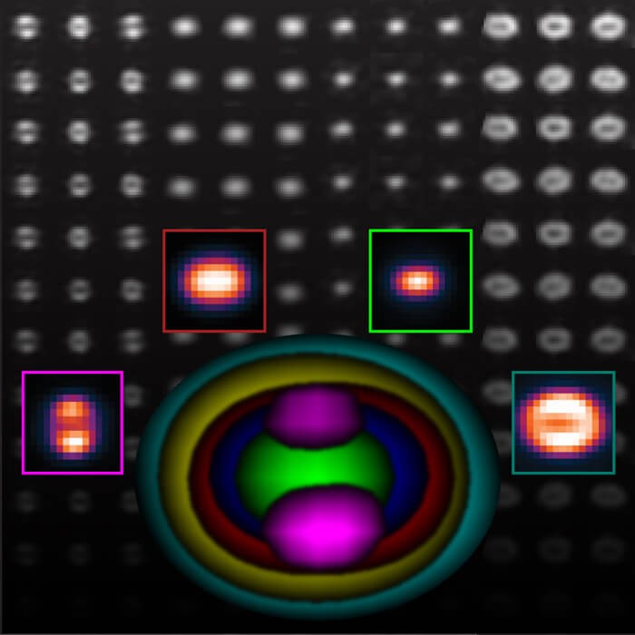
VirusMapper is an open-source software tool that enables high-resolution mapping of virus architecture using super-resolution microscopy and single particle analysis. It allows researchers to generate detailed molecular models of viruses by imaging thousands of individual virus particles labeled with fluorescent tags and averaging them together.
The key capabilities and workflow of VirusMapper are:
Segmentation - It automatically detects and segments thousands of individual fluorescently labeled virus particles from microscopy images. The particles are detected based on their shape and alignment, then cropped out and separated into individual images.
Seed selection - A subset of clearly aligned virus particle images are manually selected by the user as "seeds" to serve as templates for alignment of all the other particles. Multiple seeds representing different orientations can be selected.
Alignment and averaging - Each segmented virus particle is computationally aligned and registered to the seed templates based on rotational and translational shifts to maximize cross-correlation. Aligned particles are then averaged together, weighted by their similarity to the seeds.
Model generation - Alignment, registration and averaging are iteratively repeated using the newly generated averages as better seeds to progressively improve the final model. This allows models to be generated from very noisy individual particles.
Mapping - By labeling different virus components like outer proteins, genomes, etc. with separate fluorescent tags, models can be built independently and overlaid for multi-component mapping.
The key benefits are:
1) Molecular specificity - Unlike EM, fluorescent labeling allows specific virus proteins to be visualized
2) High resolution - Super-resolution microscopy enables sub-100nm resolution, surpassing optical diffraction limit
3) Statistical power - Thousands of single particles are analyzed compared to 10s by EM, improving accuracy
4) Context preservation - Viruses can be imaged in intact, natural states or in host cell contexts
5) Automation - VirusMapper performs automated, unbiased single particle analysis with little user input
The authors demonstrate VirusMapper by generating multi-component models of vaccinia virus architecture through SIM and STED super-resolution microscopy. They map the locations of different virus core proteins, genomes, and membranes within intact vaccinia virions. Measurements match previous EM data but also reveal new organizational details.
Additionally, by imaging cell-bound viruses, VirusMapper detects nanoscale structural changes in the virus core indicative of initial uncoating stages. This demonstrates how it can provide biological insights not accessible by EM.
VirusMapper is a powerful, open-source tool for mapping virus architecture at molecular resolution through computational processing of super-resolution microscopy images. It has the potential to uncover new structural-functional details of many viruses relevant to biomedicine and health.
Publications featuring NanoJ-VirusMapper
|
Mapping molecular complexes with super-resolution microscopy and single-particle analysis Afonso Mendes, Hannah S. Heil, Simao Coelho, Christophe Leterrier, Ricardo Henriques Review published in Open Biology, July 2022 Technologies: NanoJ-VirusMapper, Nuclear-Pores as references and ZeroCostDL4Mic () Funded by: EMBO, ERC and Wellcome Trust DOI: 10.1098/rsob.220079 |
|

|
Application of Super-Resolution and Advanced Quantitative Microscopy to the Spatio-Temporal Analysis of Influenza Virus Replication Emma Touizer, Christian Sieben, Ricardo Henriques, Mark Marsh, Romain F. Laine Review published in Viruses, February 2021 Technologies: NanoJ-VirusMapper and Super-Beacons Funded by: Wellcome Trust DOI: 10.3390/v13020233 |
|

|
Nanoscale polarization of the entry fusion complex of vaccinia virus drives efficient fusion Robert D. M. Gray, David Albrecht, Corina Beerli, Moona Huttunen, Gary H. Cohen, Ian J. White, Jemima J. Burden, Ricardo Henriques, Jason Mercer Paper published in Nature Microbiology, July 2019 Technologies: NanoJ () and NanoJ-VirusMapper Funded by: BBSRC and Wellcome Trust Blogs: Nature Microbiology Community DOI: 10.1038/s41564-019-0488-4 |
|

|
Automating multimodal microscopy with NanoJ-Fluidics Pedro Almada, Pedro M. Pereira, Siân Culley, Ghislaine Caillol, Fanny Boroni-Rueda, Christina L. Dix, Guillaume Charras, Buzz Baum, Romain F. Laine, Christophe Leterrier, Ricardo Henriques Paper published in Nature Communications, March 2019 Technologies: NanoJ (), NanoJ-Fluidics (), NanoJ-SQUIRREL (), NanoJ-SRRF () and NanoJ-VirusMapper Funded by: BBSRC and Wellcome Trust News: Technology Times, MSN, DNYUZ and Express Informer DOI: 10.1038/s41467-019-09231-9 |
|
|
NanoJ - a high-performance open-source super-resolution microscopy toolbox Romain F Laine, Kalina L Tosheva, Nils Gustafsson, Robert D M Gray, Pedro Almada, David Albrecht, Gabriel T Risa, Fredrik Hurtig, Ann-Christin Lindås, Buzz Baum, Jason Mercer, Christophe Leterrier, Pedro M Pereira, Siân Culley, Ricardo Henriques Paper published in Journal of Physics D - Applied Physics, January 2019 Technologies: CARE (), NanoJ (), NanoJ-SQUIRREL (), NanoJ-SRRF (), NanoJ-VirusMapper and QuickPALM Funded by: BBSRC and Wellcome Trust DOI: 10.1088/1361-6463/ab0261 |
|

|
Septins recognize and entrap dividing bacterial cells for delivery to lysosomes Sina Krokowski, Damian Lobato-Marquez, Arnaud Chastanet, Pedro Matos Pereira, Dimitrios Angelis, Dieter Galea, Gerald Larrouy-Maumus, Ricardo Henriques, Elias T Spiliotis, Rut Carballido-Lopez, Serge Mostowy Published in Cell host & microbe, January 2018 Technologies: NanoJ-VirusMapper Funded by: BBSRC DOI: 10.1016/j.chom.2018.11.005 |
|
|
Open-source single-particle analysis for super-resolution microscopy with VirusMapper Robert DM Gray, Jason Mercer, Ricardo Henriques Published in Journal of visualized experiments - JoVE, January 2017 Technologies: NanoJ-VirusMapper Funded by: BBSRC DOI: 10.3791/55471 |
|
__wordcloud.jpg)
|
Open-source single-particle analysis for super-resolution microscopy with VirusMapper Robert DM Gray, Jason Mercer, Ricardo Henriques Paper published in JoVE, January 2017 Technologies: NanoJ-VirusMapper Funded by: BBSRC DOI: 10.3791/55471 |
|

|
VirusMapper - open-source nanoscale mapping of viral architecture through super-resolution microscopy Robert DM Gray, Corina Beerli, Pedro Matos Pereira, Kathrin Maria Scherer, Jerzy Samolej, Christopher Karl Ernst Bleck, Jason Mercer, Ricardo Henriques Paper published in Scientific reports, January 2016 Technologies: NanoJ-VirusMapper Funded by: BBSRC DOI: 10.1038/srep29132 |
|
Funding contributing to NanoJ-VirusMapper
|
|
VirusAwareScopes - Machine Learning-Driven Adaptive Microscopy for Long-Term Viral Infection Studies Ricardo Henriques Alias: VirusAwareScopes Funded by: La Caixa Foundation - Health Research Duration: November 2025 - October 2028 |
|
|
Sub-cellular Metabolic Compartmentalization During Oocyte Development Zita Carvalho dos Santos, Ricardo Henriques, Jorge Carvalho Funded by: CZI - Measuring Metabolism Across Scales Duration: January 2024 - December 2026 |
|
|
Mapping the early stages of HIV-1 infection by live-cell 4D Super-Resolution Microscopy Hannah Heil Funded by: EMBO - Postdoctoral Fellowships Duration: September 2021 - August 2023 Publications: 5 |
|
|
Optial Biology PhD programme Michael Hausser, Ricardo Henriques, Antonella Riccio Funded by: Wellcome Trust - 4-year PhD Programme in Science Duration: August 2021 - August 2025 |
|
|
Enabling Live-Cell 4D Super-Resolution Microscopy Guided by Artificial Intelligence Ricardo Henriques Alias: SelfDriving4DSR Funded by: ERC - Consolidator Duration: July 2021 - June 2026 Publications: 38 |
|
|
Unveiling live-cell viral replication at the nanoscale Ricardo Henriques Funded by: EMBO - Installation Grant Duration: January 2021 - December 2025 Publications: 34 |
|
|
Minimal Model of Poxvirus Infection David Albrecht Funded by: Marie Curie - Postdoctoral Fellowships Duration: September 2017 - August 2019 |